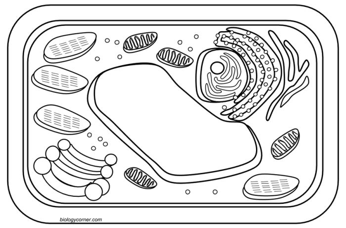
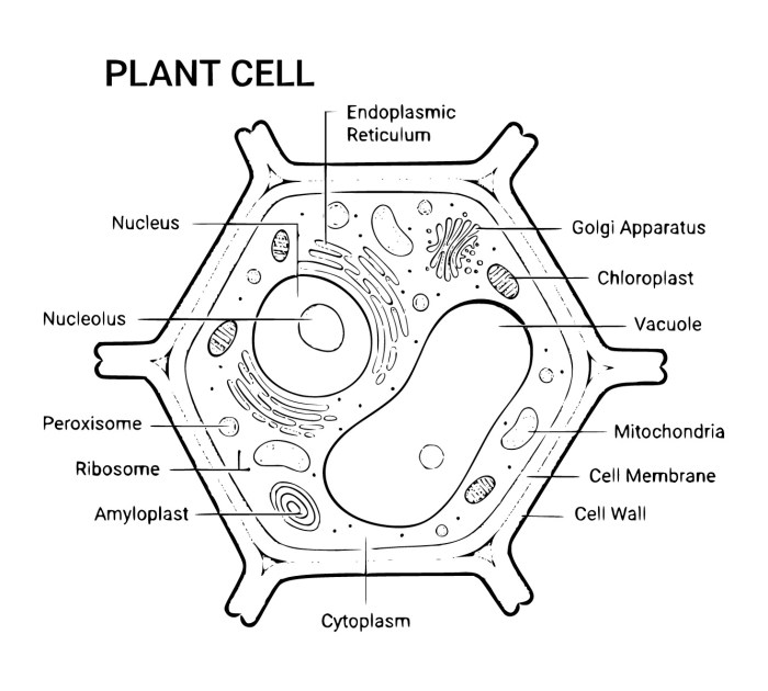
Introduction to Animal and Plant Cell Coloring Pages
Animal and plant cell coloring pages – Coloring pages depicting animal and plant cells offer a unique and engaging method for teaching fundamental biology concepts to children of various ages. The visual nature of coloring activities enhances comprehension and retention, transforming potentially abstract scientific information into a hands-on, enjoyable learning experience. By coloring the different organelles and structures, students actively engage with the material, fostering a deeper understanding of cell function and structure than passive learning methods might provide.The key difference between animal and plant cells lies in the presence of specific organelles unique to plant cells.
These differences should be clearly illustrated in coloring pages to emphasize the distinct characteristics of each cell type. Accurate representation of these differences is crucial for effective learning and prevents misconceptions from developing. Highlighting these differences promotes a comparative understanding, allowing students to readily identify and differentiate between the two cell types.
Age-Appropriate Coloring Page Designs
The design and complexity of coloring pages should be tailored to the age and developmental stage of the child. Preschoolers benefit from simplified diagrams with large, clearly defined shapes and limited details. Elementary school children can handle slightly more complex designs, incorporating more organelles and labeling opportunities. Middle school students can benefit from more detailed diagrams, possibly including three-dimensional representations or cross-sections, allowing for a more advanced understanding of cellular processes.
For example, a preschool coloring page might simply depict a circle for the cell membrane, with a few larger, differently colored shapes representing the nucleus and cytoplasm. An elementary school page could add organelles like mitochondria and vacuoles, with simple labels. A middle school page could depict a more detailed structure, potentially including the endoplasmic reticulum, Golgi apparatus, and chloroplasts (in the case of plant cells), with clear labeling and perhaps a simplified diagram showing the functions of each organelle.
Preschool-aged children’s coloring pages should use bold Artikels and large, easily colored shapes, focusing on the basic components of the cell: the cell membrane, nucleus, and cytoplasm. These simplified representations emphasize the overall structure without overwhelming the child with excessive detail. The colors used should be bright and engaging to maintain the child’s interest.
Elementary school-aged children can handle slightly more intricate designs. The coloring page can include additional organelles such as mitochondria, vacuoles, and, in the case of plant cells, chloroplasts. Simple labels can be incorporated to help children learn the names of these components. The use of different colors for each organelle enhances differentiation and understanding.
Middle school-aged children can be presented with more complex diagrams. These could include three-dimensional representations of the cells, showing the spatial relationships between organelles. Cross-sections can be incorporated to illustrate the internal structures more clearly. More detailed labels and explanations of the function of each organelle can also be included to facilitate a deeper understanding of cellular processes.
The level of detail can also include depictions of cell processes such as photosynthesis or cellular respiration.
Educational Applications and Activities
Coloring pages depicting animal and plant cells offer a unique and engaging approach to teaching fundamental concepts in cell biology. The visual nature of the activity makes complex structures more accessible to students of all ages and learning styles, fostering a deeper understanding of cell components and their functions. These activities can be integrated into various educational settings, from elementary school classrooms to high school biology labs, and even used as supplemental learning tools for homeschooling.The act of coloring itself facilitates active learning and improves memory retention.
Animal and plant cell coloring pages offer a unique educational opportunity, allowing students to visualize the intricate structures of life. This microscopic exploration contrasts sharply with the macroscopic world depicted in coloring in pages of animals , which focus on the external forms and characteristics of entire organisms. Returning to the cellular level, these coloring pages provide a valuable tool for understanding the fundamental building blocks of both plant and animal life.
By meticulously coloring the different organelles and structures, students actively engage with the material, reinforcing their knowledge and improving recall. This hands-on approach transforms a potentially abstract topic into a concrete and memorable experience, significantly impacting learning outcomes. Furthermore, coloring pages can be adapted to cater to different learning styles, making them a valuable tool for inclusive education.
Activities Enhancing Cell Structure and Function Understanding
Coloring pages provide a foundation for a range of activities that extend beyond simple coloring. Students can label the various organelles after coloring, creating a personalized cell diagram. This activity reinforces the names and locations of structures like the nucleus, mitochondria, chloroplasts (in plant cells), and cell walls. Furthermore, teachers can incorporate interactive exercises where students match organelle names to their functions, strengthening their understanding of cellular processes.
Comparative analysis activities, where students compare and contrast animal and plant cells by identifying similarities and differences in their structures and functions, can also be incorporated. For example, a teacher could ask students to explain why plant cells have chloroplasts but animal cells do not, prompting deeper engagement with photosynthesis and energy production. Finally, creating a 3D model of a cell after coloring can provide a tactile learning experience that solidifies understanding of spatial relationships within the cell.
Reinforcing Concepts Learned in Science Classes
Coloring pages serve as effective reinforcement tools after a lesson on cell structure and function. They provide a visual summary of the key concepts discussed in class, allowing students to revisit and solidify their understanding. For example, after a lecture on the role of mitochondria in cellular respiration, students can color the mitochondria in their coloring pages, actively connecting the visual representation to the learned concept.
Teachers can assign targeted coloring activities based on specific lesson topics, turning them into a formative assessment tool. For instance, students can focus on coloring only the organelles related to protein synthesis after learning about ribosomes and the endoplasmic reticulum. This focused approach helps reinforce specific aspects of the lesson. Additionally, differentiated instruction can be implemented by providing students with different levels of complexity in their coloring pages, catering to diverse learning needs and paces.
Assessing Student Understanding of Cell Biology, Animal and plant cell coloring pages
Coloring pages, when combined with other assessment methods, can provide valuable insights into student understanding. Teachers can assess comprehension by asking students to label the colored organelles, explain the functions of specific structures, or compare and contrast the structures of animal and plant cells based on their completed coloring pages. This method offers a low-stakes assessment opportunity that can reveal misconceptions and inform future teaching strategies.
Open-ended questions, such as asking students to explain the importance of the cell membrane in maintaining homeostasis, can be incorporated to assess deeper understanding beyond simple identification. The teacher can also use the coloring pages as a starting point for discussions about cell biology, fostering a collaborative learning environment where students can share their understanding and learn from each other.
Finally, the completed coloring pages can be used as part of a portfolio assessment, demonstrating student progress and understanding of cell biology over time.
Illustrative Examples

The following descriptions provide detailed visualizations of key organelles within animal and plant cells, highlighting their structural features and functional roles. These examples serve to enhance understanding of the differences and similarities between these two fundamental cell types. Visualizing these structures aids in grasping the complexities of cellular processes.
Mitochondrion in an Animal Cell
The mitochondrion, often referred to as the “powerhouse of the cell,” is a double-membraned organelle crucial for cellular respiration. Imagine a bean-shaped structure with a highly folded inner membrane. This inner membrane, known as the cristae, significantly increases the surface area available for the electron transport chain, a key step in ATP (adenosine triphosphate) production. The space between the inner and outer membranes is called the intermembrane space, while the space enclosed by the inner membrane is the mitochondrial matrix.
Within the matrix, the citric acid cycle (Krebs cycle) takes place, generating high-energy electron carriers that fuel the electron transport chain on the cristae. The resulting ATP molecules provide the energy necessary for various cellular processes. The outer membrane is relatively smooth and permeable, allowing the passage of small molecules. The mitochondrion also possesses its own DNA (mtDNA) and ribosomes, suggesting an endosymbiotic origin.
Chloroplast in a Plant Cell
The chloroplast, unique to plant cells and some protists, is the site of photosynthesis. Picture a lens-shaped organelle containing a complex internal structure. Its defining feature is a system of interconnected, flattened membrane sacs called thylakoids. These thylakoids are stacked into structures called grana, which are interconnected by stroma lamellae. The thylakoid membranes house the chlorophyll and other pigments crucial for capturing light energy.
The space inside the thylakoids is called the thylakoid lumen. Surrounding the thylakoids is the stroma, a fluid-filled region where the Calvin cycle takes place, converting carbon dioxide into sugars using the energy captured during the light-dependent reactions in the thylakoids. Like mitochondria, chloroplasts also possess their own DNA and ribosomes, supporting the endosymbiotic theory.
Cell Wall, Vacuole, and Nucleus: Plant vs. Animal Cells
The cell wall, vacuole, and nucleus exhibit significant differences in plant and animal cells.The cell wall, a rigid outer layer, is present only in plant cells. Imagine a strong, protective outer shell composed primarily of cellulose. It provides structural support and protection, maintaining cell shape and preventing excessive water uptake. Animal cells lack this rigid outer layer, relying instead on their cell membrane for structural integrity.The vacuole, a large, fluid-filled sac, is considerably larger in plant cells than in animal cells.
In plant cells, visualize a central vacuole occupying a significant portion of the cell’s volume. It stores water, nutrients, and waste products, contributing to turgor pressure that maintains cell shape and rigidity. Animal cells may contain smaller vacuoles involved in various functions, such as waste removal, but they do not typically dominate the cell’s interior like the central vacuole in plant cells.The nucleus, the control center of the cell, is present in both plant and animal cells.
Imagine a spherical structure enclosed by a double membrane, the nuclear envelope. This envelope contains nuclear pores that regulate the transport of molecules between the nucleus and the cytoplasm. Inside the nucleus, the genetic material, DNA, is organized into chromosomes. While the basic structure of the nucleus is similar in both cell types, the size and relative position within the cell might vary slightly depending on the cell type and its stage of the cell cycle.
The nucleolus, a dense region within the nucleus, is involved in ribosome synthesis, and is present in both plant and animal cell nuclei.
Accessibility and Inclusivity

Creating engaging and educational coloring pages requires careful consideration of accessibility and inclusivity to ensure all students can participate and benefit. This involves adapting the materials for students with visual impairments and representing the diversity of the natural world accurately and respectfully. Furthermore, design choices must cater to a broad spectrum of learning styles and abilities.Designing coloring pages that are accessible and inclusive requires a multifaceted approach.
This includes considering the needs of students with visual impairments, ensuring diverse representation of flora and fauna, and creating engaging activities suitable for a range of learning styles and abilities.
Coloring Pages for Visually Impaired Students
Adapting coloring pages for students with visual impairments requires the incorporation of tactile elements and alternative formats. One strategy is to create raised-line drawings using materials like thick paper, cardboard, or textured fabrics. These raised lines allow students to trace the Artikels of the cells and their components, gaining a kinesthetic understanding of their shapes and relationships. Another approach is to provide large-print versions of the coloring pages with bold Artikels and contrasting colors.
Furthermore, the use of Braille labels to identify the different parts of the cells can enhance the learning experience for visually impaired students. Finally, audio descriptions of the cell structures and their functions can be provided alongside the coloring pages, offering an auditory learning experience.
Diverse Representation of Animals and Plants
Incorporating diverse representations of animals and plants is crucial for creating inclusive coloring pages. This means showcasing a wide range of species, reflecting the biodiversity of the planet and avoiding stereotypical or limited representations. For example, instead of only depicting common domesticated animals like cats and dogs, the coloring pages could include less frequently represented animals from various ecosystems, such as a pangolin from Africa, a koala from Australia, or a snow leopard from the Himalayas.
Similarly, plant diversity should be represented by showcasing various types of trees, flowers, and other plant life, including those from different regions and climates. This approach helps students appreciate the vastness and beauty of the natural world and fosters respect for biodiversity. Including plants that are relevant to different cultures and their traditions can further enhance inclusivity.
For example, a rice plant would be meaningful for students from Asian cultures, while a corn plant could be important for those from the Americas.
Engaging and Appealing Designs for Diverse Learners
Creating engaging and appealing coloring pages for a wide range of learners necessitates considering different learning styles and preferences. Some students may benefit from simpler designs with clear Artikels and large spaces for coloring, while others may prefer more complex and detailed drawings that challenge their fine motor skills. Incorporating a variety of textures and patterns within the drawings can also add visual interest and cater to different preferences.
Providing choices in terms of coloring media—crayons, markers, colored pencils, paints—allows students to use their preferred tools, enhancing their engagement and creativity. Furthermore, integrating interactive elements, such as simple puzzles or matching games related to the cell structures, can make the coloring pages more stimulating and effective for a wider range of learners. The inclusion of fun facts or trivia related to the animals and plants depicted can also enhance the educational value and appeal of the coloring pages.

